
किपा:Tubular adenoma 2 intermed mag.jpg
Size of this preview: ८०० × ५३३ pixels. Other resolutions: ३२० × २१३ pixels | ६४० × ४२७ pixels | १,०२४ × ६८३ pixels | १,२८० × ८५३ pixels | २,५६० × १,७०७ pixels | ४,२७२ × २,८४८ pixels.
Original file (४,२७२ × २,८४८ pixels, file size: ४.४७ MB, MIME type: image/jpeg)
Summary
| Description |
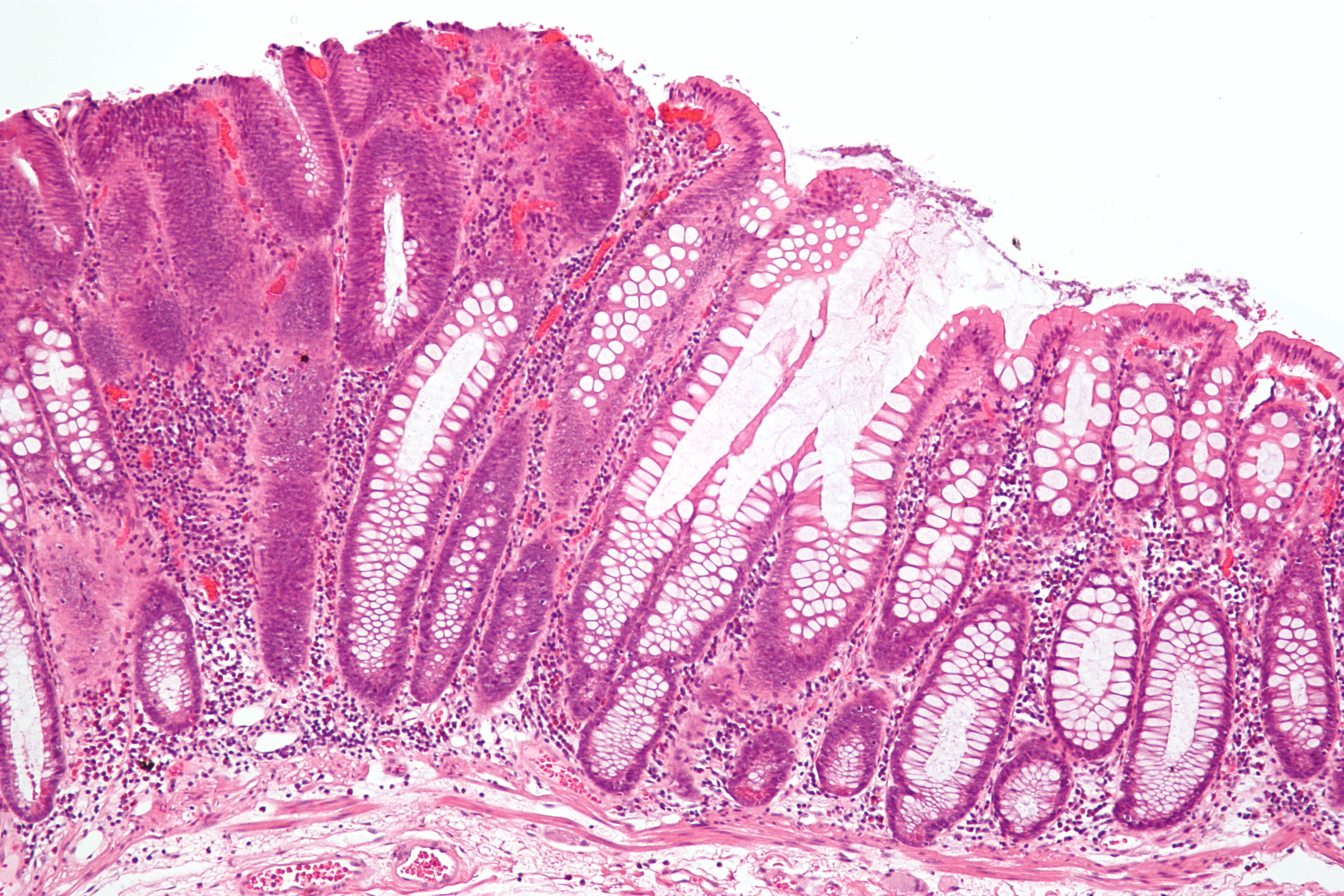
English: Intermediate micrograph of a colorectal tubular adenoma without high grade dysplasia. H&E stain.
The lesional tissue, i.e. dysplastic epithelium, is seen on the left of the image and characterized by:
Normal colonic type epithelium is seen on the right of the image and characterized by small round nuclei and abundant goblet cells. Related imagesThe same case:
Another case:
|
| Source | Own work |
| Author | Nephron |


Licensing
I, the copyright holder of this work, hereby publish it under the following licenses:
This file is licensed under the Creative Commons Attribution-Share Alike 3.0 Unported license.
- You are free:
- to share – to copy, distribute and transmit the work
- to remix – to adapt the work
- Under the following conditions:
- attribution – You must give appropriate credit, provide a link to the license, and indicate if changes were made. You may do so in any reasonable manner, but not in any way that suggests the licensor endorses you or your use.
- share alike – If you remix, transform, or build upon the material, you must distribute your contributions under the same or compatible license as the original.

|
Permission is granted to copy, distribute and/or modify this document under the terms of the GNU Free Documentation License, Version 1.2 or any later version published by the Free Software Foundation; with no Invariant Sections, no Front-Cover Texts, and no Back-Cover Texts. A copy of the license is included in the section entitled GNU Free Documentation License. |
You may select the license of your choice.
File history
Click on a date/time to view the file as it appeared at that time.
| Date/Time | Thumbnail | Dimensions | छ्य्लामि | Comment | |
|---|---|---|---|---|---|
| current | ०१:०९, २७ अक्टोबर २००९ | | ४,२७२ × २,८४८ (४.४७ MB) | Nephron | {{Information |Description={{en|1=Intermediate micrograph of a colorectal '''tubular adenoma''' without high grade dysplasia. H&E stain. The lesional tissue, i.e. dysplastic epithelium, is seen on the lef |
File usage
The following page uses this file:
Global file usage
The following other wikis use this file:
- Usage on ar.wikipedia.org
- Usage on de.wikibooks.org
- Usage on en.wikipedia.org
- Usage on es.wikipedia.org
- Usage on fa.wikipedia.org
- Usage on fr.wikipedia.org
- Usage on gl.wikipedia.org
- Usage on he.wikipedia.org
- Usage on hi.wikipedia.org
- Usage on id.wikipedia.org
- Usage on it.wikipedia.org
- Usage on ja.wikipedia.org
- Usage on ko.wikipedia.org
- Usage on la.wikipedia.org
- Usage on ml.wikipedia.org
- Usage on ms.wikipedia.org
- Usage on pt.wikipedia.org
- Usage on simple.wikipedia.org
- Usage on sr.wikipedia.org
- Usage on sv.wikipedia.org
- Usage on te.wikipedia.org
- Usage on th.wikipedia.org
- Usage on uk.wikipedia.org
- Usage on zh.wikipedia.org